Professor Lee’s diagnosis pathway
A clear pathway to understanding your symptoms.
Professor Paul Lee brings your symptoms, examination and investigations together to identify the cause of exercise-related lower-leg pain and plan treatment. His pathway combines contrast MRI, exercise-related CK blood tests, body measurements and ExtPress™ compartment testing, selected for your clinical needs.
Start with the person
Your symptoms, movement and measurements
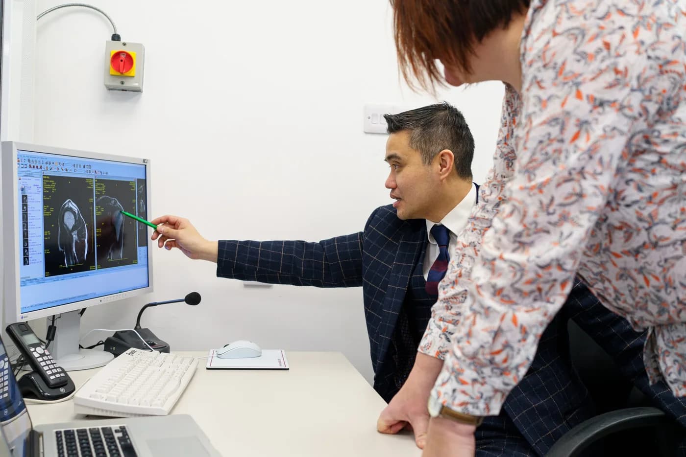
The assessment starts with the activity that brings on pain, its location and how it settles. Professor Lee examines your legs and reviews your movement. Height, the thigh-to-cleft length ratio and maximum calf circumference are recorded as part of the physical assessment. Supervised exercise helps the team observe the symptoms you experience during activity.
Professor Lee’s pathway
Findings brought together into a clear diagnosis and treatment recommendation.
Exercise. Measurement. Clinical insight.
ExtPress™: measuring compartment pressure
ExtPress™ measures pressure within the muscle compartments around exercise. It gives Professor Lee direct information about the compartments involved and their response to activity. He interprets the readings with your symptoms and the exercise and measurement timings. Published protocols vary; the 2022 expert consensus did not agree on a universal pressure cut-off.
NHS: compartment syndrome · Vogels et al. (2022): clinical consensus on lower-leg CECS
Contrast MRI
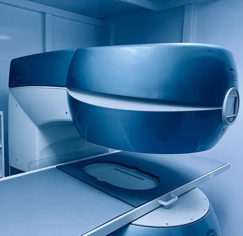
Contrast MRI: looking for abnormalities
Professor Lee’s pathway uses contrast MRI to look for abnormalities in the lower leg and investigate other explanations for your symptoms. The imaging team reviews your medical history and contrast suitability before the scan. Human studies also support exercise MRI as a way to examine muscle changes associated with CECS; the scan requested for you is tailored to the clinical question.
Ringler et al. (2013): exercise MRI validation in 76 patients · NHS: compartment syndrome
“What are we looking for, and how would the answer change my care?”A question you can ask
Muscle response before and after exercise
Before-and-after exercise CK blood tests
Blood samples before and after exercise measure creatine kinase (CK), an enzyme associated with muscle activity and injury. Professor Lee reviews that response alongside the examination, MRI and pressure findings. These findings are assessed together to reach a clinical diagnosis and recommend the next step. Bring previous reports and scan access details so the team can build on investigations you have already had.
- Before exercise
A baseline CK blood sample.
- After exercise
A second sample to review the CK response.
- Findings together
CK, MRI, measurements, symptoms and pressure.
Common questions
Will everyone need a pressure test?
Not necessarily. Test selection depends on your symptoms, examination, previous investigations and the decision the clinician needs to make.
Should I bring my previous test results?
Yes, if available. Bring reports, scan access details and any pressure-test record, including the activity and timing of measurements. This can help the clinician interpret previous findings.